Case reports
← vista completaPublished on May 13, 2015 | http://doi.org/10.5867/medwave.2015.04.6137
Sacrococcygeal teratoma: case report
Teratoma sacrococcígeo: presentación de un caso
Abstract
We present a male newborn child with a sacrococcygeal mass who was sent to clinic 46 of the Mexican Social Security Institute located in Gomez Palacio, Durango, Mexico for pediatric/neonatal surgical resolution. The mass was detected on gestation week 24 in the sacrococcygeal area and was initially interpreted as a myelomeningocele. On gestation week 32, the mass had grown, so the diagnosis of cystic hygroma was posed. The child was born at 38 weeks of gestational age with a large tumor in the sacrococcygeal area. Images were obtained, and tumor resection was performed without complications. Pathologic examination confirmed the diagnosis of sacrococcygeal teratoma. The postoperative course was uneventful and there were no further complications.
Introduction
Sacrococcygeal teratoma is a neoplasia which despite its low frequency, is one of the most common ones in neonates [1]. Unlike in adults, fetal teratomas mostly develop at sacrococcygeal level. They are formed of various types of tissue derived from at least two, or the three embryonic layers [2]. These tumors may get enormous dimensions and contain large blood vessels that provoke blood depriving to the developing fetus.
The best way to treat this malignancy, is through complete resection and a histopathological study of it. With this approach it is possible to prevent late complications such as coagulopathy, necrosis by compression, infections or intratumoral hemorrhage [3].
The differential diagnosis must be made with the myelomeningocele - entity that more frequently leads to confusion with other diseases such as lipomas, hemangiomas, pilonidal cyst and epidermoid cyst. Furthermore, the teratoma must be differentiated according to its location, between the coccyx and the anus, from others that are located behind the sacrum and even in the intrapelvic region [4].
The availability of obstetric ultrasound has been a great breakthrough that has allowed the prenatal diagnosis of many of these lesions. With it, timely planning, study and multidisciplinary treatment of patients can be achieved [5], as summarized in the case presented below.
Presentation of the case
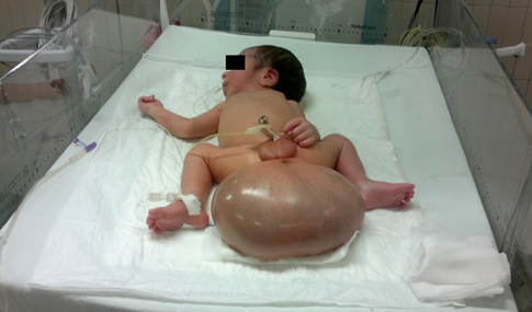
Male newborn, 18 year old mother. Product of gestation: 1. A Rh positive blood type. Normal evolution of pregnancy, prenatal care from the beginning, attended eleven medical consultations. Presence of tumor mass in sacrococcygeal region was detected by ultrasound since 24 weeks of pregnancy. At first it was categorized as probable myelomeningocele, consequently, the patient was sent for perinatal handling to the third level of care at clinic 16 of the Mexican Social Security Institute in Torreón Coahuila, Mexico. A control ultrasound was performed at 32 weeks of pregnancy in which tumor volume increased with a probable diagnosis of cystic hygroma. It was scheduled for cesarean section at 38 weeks of gestation and a single live male product is obtained, with Apgar 8-9, Silverman Anderson 0, Capurro test : 38 weeks of gestation, weight 4500 g.
Physical examination: normal head; cardiopulmonary system not involved; soft, depressible abdomen, without organ enlargement. One tumor with about 1 kg of weight, 16.5x8.5x12 cm dimensions and liquid appearance in approximately 50% of its size can be appreciated in the sacrococcygeal region. (Figure 1)
 Full size
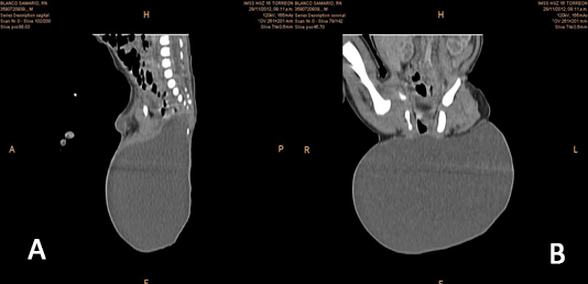
 A simple and contrasted axial computerized tomography of pelvis is performed. A proper aligned complete lumbosacral spine is observed in the tomography. The bone structure of the pelvis has no alterations. Bladder with little repletion and unaltered. Distal rectum with anterior orientation. Suggestive image of anal opening can be seen in previous perianal region. Testicles and penis with no alterations. Soft tissues in perianal region show cystic aspect in a large image with 16.5 cm of transversal diameter by 8.5 cm anteroposterior and 12 cm longitudinal, with presence of three calcium images on its wall.
Through the administration of contrasting agent, reinforcement of the wall is observed, as well as a thin septum in its posterior wall (Figure 2A and B).
 Full size
 More images can be observed at the following link.
Abdominal ultrasound was reported unaltered.
After his birth, he was sent to the clinic 46 of the Mexican Social Security Institute of Gomez Palacio, Durango, Mexico, for its handling and neonatology pediatric surgery.
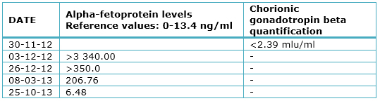
In pediatric surgery is scheduled for resection of tumor, previous indication of tumor markers analysis. These markers report quantification of gonadotropin chorionic human β: 2.39 mIU / ml within normal parameters; alpha-fetoprotein: 3,340 ng / ml (reference: 0 to 13.4) and normal preoperative tests. Surgery is performed seven days after birth.
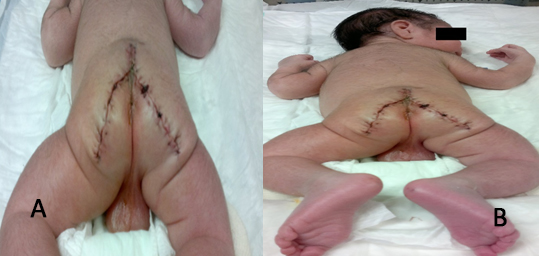
Dissection by planes is performed following tumor planes with nutritious vessels ligation and hemostasis with cautery, rectum is located prior catheter with Hegar 7 Fr. Adhered rectal tumor but not invasive is dissected and coccyx is resected, tying median sacral artery, besides resecting one small percentage of the tumor at presacal level. Drain suction is placed and it is closed by layers (Figure 3 A and B).
 Full size
 Findings
A 18x14x10 cm encapsulated tumor was found Altman I. Approximately 80% of its composition is of citrine cystic type and 20% solid. A 100% noninvasive, resection of the tumor that was attached to the rectum was performed, with a minimal percentage of presacral portion; coccyx was also resected.
Pathology report
Macroscopic description: irregularly ovoid specimen is received measuring 16 cm in diameter. The surface shows areas coated by a brown clear and smooth epidermis and bloody appearance areas. When cutting is soft. A unilocular cystic lesion is observed. The walls have a maximum thickness of 0.2 cm. Internal face is smooth light brown, shiny, with reticular appearance areas. A mamelon is seen in one of the poles measuring 4x2.8 cm the area is light brown with congested areas. When cutting is soft with hard areas. The surface is light with chondroid appearance areas.
Microscopic description: malignancy of germinal origin is identified. The cyst wall consists of dense connective tissue coated in the inside by simple squamous epithelium, with no evident alterations. Underlying skin without histological alterations observed. In one pole, a mamelon can be observed in which remnants of glial and choroid tissue, skin, epithelium of respiratory aspect, cartilage, and congestive vessels are identified. From a central point, bone with very little presence of bone marrow is presented. All components are mature and no malignancy data is observed. No percentage of neuroepithelium is presented.
During patient follow-up, alpha-fetoprotein levels decreased to normal values until now. (Table 1)
 Full size
 Discussion
This case is presented because of its low incidence and its rarity. About 3,000 births occur annually at the clinic number 46 of the Mexican Social Security Institute in Gomez Palacio, Durango. We expect to find a case of this nature every 13 to 15 years.
Sacrococcygeal teratomas are extragonadal neoplasms arising in the presacral area. They have an incidence of one per 40,000 live births, and a prevalence of one in 21,000 births. Although sacrococcygeal teratoma is a rare tumor, it is the most common malignancy of germ cells in newborns and children under two years. Its presentation may be forming large cysts or as solid mass. They are usually located in the midline of the body. In general, the order of frequency regarding to its location is as follows: sacrococcygeal, gonadal, retroperitoneal, cervical, mediastinal, oropharyngeal and other (gastric, hepatic, intracranial). It predominates in females, but in males malignancy is more recurrent [6],[7],[8].
The term "teratoma," derives from the Greek word "teraton" meaning monster. In 1869 Virchow applied this term to a tumor originating in the sacrococcygeal region [9].
Teratomas are formed by multiple foreign tissues to the organ or place where they are produced. Most lesions occur in the neonatal period and may be benign or malignant, cystic or solid. Although teratomas are sometimes defined by having the three embryonic layers (endoderm, mesoderm and ectoderm), recent classifications include the monodermal type [8].
Sacrococcygeal teratoma can grow to huge dimensions, causing complications related to mass effect such as distortion of the anatomy of the pelvis and sacrum, bladder obstruction and dystocia [6],[10].
The Section on surgery of The American Academy of Pediatrics proposed a useful clinical postnatal classification system, that describes four types of sacrococcygeal teratoma according to their location [11]:
- Type I: predominantly external, with minimum presacral component. It is the most common and the least malignant (45.8%).
- Type II: External with significant intrapelvic component
- Type III: External with pelvic mass. In this one, it predominates an extension through the abdomen.
- Type IV: It is presacral without external presentation or significant pelvic extension. It is the most malignant.
As for their tissue composition, there are three histological main types [12],[13]:
- Mature: well differentiated tissues such as brain, skin and bones.
- Immature: neuroplia, neural tube-like structures in addition to mature components. They have high incidence of malignancy. There are four categories depending on the amount of immature tissue present and the mitotic activity degree.
- Teratoma with malignant components: this teratoma contains one or more malignant germ cell tumors. For example choriocarcinoma, germinoma, embryonal carcinoma, endodermal sinus tumor; in addition to mature or immature tissue.
Two major problems remain to be solved: longterm functional sequelae (fecal and urological) in addition to recurrence [14],[15],[16],[17],[18],[19]. Tuladhar et al. [20] mentioned in a review in 2000, that in most publications recurrence rate of sacrococcygeal teratomas varies between 7.5 and 22% of patients. On the other hand, in a series of 20 cases published in Mexico in 2003 by Gutiérrez Ureña et al. [4], postsurgical evolution was satisfactory and cases remained free of disease during 36 months follow up. Some possible causal factors for recurrence include incomplete resection with microscopic residues, non all coccyx resection and tumor spread.
Diagnosis as a large mass protruding from the sacral /buttock is often performed in utero by ultrasound. Other findings include erosion of a vertebral body or soft tissue mass with areas of calcification in radiography [21],[22].
It is important to identify whether the lesions are cystic or not, since cystic lesions have a better prognosis than solid ones, women also have a better prognosis than men. Lesions diagnosed at two months of age, are more likely to contain malignant tissue. Sacrococcygeal teratomas tend to metastasize to the liver, lungs and lymph nodes. Recurrence has been found after 40 years of resection, which may be announced by alpha-fetoprotein levels. Not only is ultrasound useful for the diagnosis, but it also can be used to monitor tumor progression, detect complications and establish management [21].
Although the majority of the cases are benign, sacrococcygeal teratomas are linked to high morbidity and mortality from preterm deliveries, along with complications such as malignant invasion, tumor hemorrhage, obstruction of umbilical flow and high output heart failure, [23]. Death happens primarily in fetuses with solid and highly vascularized fast growing teratomas that can lead to high output cardiac insufficiency. This happens because the tumor acts as a large arteriovenous malformation [24]. Because some of these complications can be prenatally detected and treated appropriately, the prenatal diagnosis of sacrococcygeal teratomas is very important [25],[26].
The main treatment for sacrococcygeal teratoma, regardless of histological type, is complete resection of the tumor and the coccyx [27]. If this procedure is not performed, the risk of recurrence is extremely high. In patients with mature sacrococcygeal teratoma, the only recommended treatment is surgery. In immature sacrococcygeal teratoma, the treatment includes surgery (removal of the sacrum and coccyx), followed by observation. Sometimes a mature or immature teratoma also contains malignant cells. The teratoma and malignant cells may need to be treated differently. Regular follow-up examinations are performed by imaging procedures and tumor marker tests of alpha-fetoprotein [27].
The sacrococcygeal teratoma has a relapse rate of about 4% (39 months follow- up) after resection and a minimum three-year follow-up is set [20],[22]. There is little evidence to provide guidance on follow-up care for children with sacrococcygeal teratomas. The following tests and procedures may be performed at the physician discretion, when tumor markers are elevated at diagnosis moment: alpha-fetoprotein and human chorionic gonadotropin β. Both markers should be monitored monthly during six months (period of highest risk), and then every three months until completing three years as recommended by the National Cancer Institute, National Institutes of Health in the United States [15],[27].
Tumor markers
Yolk sac tumors produce alpha-fetoprotein, while germinomas (seminomas and dysgerminomas) and especially choriocarcinomas produce human chorionic gonadotropin β, resulting in elevated serum levels of these substances. Most children with malignant sacrococcygeal teratomas will have a component of yolk sac tumor, along with elevations in concentrations of alpha-fetoprotein, which are monitored in series during treatment to help assess their response. Benign teratomas and immature teratomas may produce small elevations of alpha-fetoprotein and human chorionic gonadotropin β [28],[29],[30],[31].
Conclusions
We can claim that sacrococcygeal teratomas are rare tumors in Mexico. They originate as a remnant of the primitive streak and may present with any histological type of the three germ layers. In the majority of the cases they are benign tumors; the risk of recurrence or malignant transformation is low. It is important to recognize the existence of this pathology in order to have the clinical expertise that offers timely diagnosis, an appropriate and multidisciplinary treatment. Monitoring with alpha-fetoprotein and ultrasound is a key to detect recurrence or postoperative complications.
Finally, reviewing the literature and researching these isolated cases that present at the second level of care, is probably the best tool to keep health professionals up to date and help them provide better care to their patients.
Notes
Readers can request more images by directly contacting the author responsible for the publication.
From the editor
This article was originally submitted in Spanish and was translated into English by the authors. The Journal has not copyedited this version.
Ethical issues
The Journal certifies that this study has been approved by the Local Committee for Research and Ethics in Health Research at the Mexican Social Security Institute. This committee refers that ethical aspects about information management and procedures were respected and it has evidence of the patient's consent to publish his case.
Conflicts of interest statement.
The authors have completed the conflict of interests declaration form from the ICMJE, which has been translated into Spanish by Medwave, and they declare that they have not received any funding whatsoever to write this article, nor have they any conflict of interests with the matter dealt herein. Forms can be requested to the responsible author or the editorial direction of the Journal.
