Case reports
← vista completaPublished on November 9, 2016 | http://doi.org/10.5867/medwave.2016.10.6598
Tinea incognito due to Trichophyton mentagrophytes: case report
Tiña incógnita debida a Trichophyton mentagrophytes: reporte de caso
Abstract
Tineas are frequent infections caused by dermatophytes that are able to invade keratinized tissue, causing rounded, erythematous, scaly lesions. Nonetheless, in tinea incognito the lesions are modified because of inappropriate use of topical corticosteroids or calcineurin inhibitors, making it difficult to diagnose. We present a case of a 12-year-old male child that presents with erythematous lesions on the right eyebrow, which at first was diagnosed as a contact dermatitis, so corticosteroids were indicated. The lesions became more inflammatory and a fungus culture was requested, which was positive for Tricophyton mentagrophytes. The conclusion of this report is that tineas can mimic other dermatologic conditions therefore caution should be taken when prescribing topical corticosteroids or calcineurin inhibitors without diagnostic certainty because if lesions are caused by tinea, diagnosis and treatment may become more difficult.
Introduction
Dermatophytosis, ringworm or tinea is an infection, frequently observed in children and adults every day. It is caused by a group of fungi called dermatophytes, which are able to invade keratinized tissue, such as the stratum corneum of the skin, hair and nails. The consequent lesions are typically erythematous, desquamative, well delimited annular plaques, with an active edge of scaly, microvesicular, centrifugal growth, associated to a paler center [1].
This “typical” clinical presentation may vary, either for characteristics of the dermatophyte itself, for example, its invasion capacity; physiopathologic conditions of the patient, such as immunosuppression, inflammatory responses of the skin, age extremes and skin barrier alterations; or for environmental conditions, as sun exposure [2]. In this context, ringworm can mimic other cutaneous disorders as rosacea, psoriasiform eruptions, seborrheic dermatitis, lupus erythematosus, atopic dermatitis, contact dermatitis, bacterial infections, herpetic infections, granuloma annulare, erythema annulare centrifugum, pityriasis rosea, sarcoidosis, Hansen’s disease and urticaria, mainly. Furthermore, there are other multiple causes for annular lesions that can mimic a dermatophyte infection, leading to incorrect diagnosis and treatment and unexpected results [3],[4],[5].
In that regard, tinea incognito is described as a dermatophyte infection in which typical clinical presentation has been modified due to the inappropriate use of topical corticosteroids or calcineurin inhibitors, normally because of an incorrect diagnosis or the patient’s self-medication [1].
Tinea incognito is a low frequency entity. Its global prevalence has not been determined, although it corresponds to less than 1% of dermatophytoses [6]. It is important to note that this pathology is preventable if there is awareness of it before prescribing any treatment with topical corticosteroids or immunosuppresses in lesions that may correspond to a dermatophyte infection.
This case of tinea incognito is presented aiming to provide knowledge regarding preventive measures to be considered at the beginning and during treatment with topical corticosteroids, even when the lesion seems “typical” of a certain cutaneous pathology. Particularly, our case mimics a contact dermatitis, which normally would be treated with topical corticosteroids. Therefore, even if ringworm diagnosis is relatively easy, this demonstrates it can become an important diagnosis challenge due to variations on its clinical presentation in some cases.
Clinical case
The patient is a 12-year-old male with a history of a controlled atopic dermatitis under regular treatment. He is an athlete, who practices motocross enduro.
He presents a three-week history of pruritic lesions located in his face, particularly in the right periocular region. For this reason, the patient schedules an appointment with a health care provider, where a severe dermatitis was diagnosed and a moderate potency topical corticosteroid was prescribed. The patient evolved favorably during the first three days of treatment.
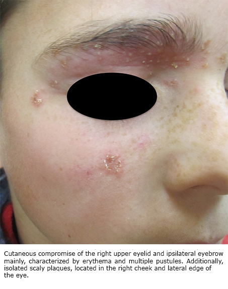
However, a week later, he attends to our medical center because the lesions had reappeared, but this time they were more inflammatory and erythematous. It also stood out that there were some pustules located mainly in the right upper eyelid and ipsilateral ciliary edge (Figure 1).
Considering the use of a helmet and goggles (friction), it was interpreted as a secondary folliculitis by friction and, additionally, because of the contact with dirt, a direct mycological examination and a basic culture were requested. Both exams were negative, so a new fungal culture was requested but this time, with samples from pilous follicles specifically. The result was positive for Trichophyton mentagrophytes. The patient had already started an empirical treatment with itraconazole, 100 mg daily, evolving favorably the first week of treatment. Initially, due to the strongly inflammatory lesions, it was decided to prescribe a less potent topical corticosteroid just for three days. Finally, the patient completed a month of itraconazole, with no complications or relapses, and after two months of the beginning of the treatment a new fungus culture was requested, resulting negative.
 Full size
 Discussion
The term “tinea incognito” is used to describe a dermatophytosis that has been clinically modified by the inappropriate use of topical corticosteroids or calcineurin inhibitors. It does not have predilection for gender or age and it is observed more frequently in the trunk (30.4%), followed by face (24.4%), feet (13.8%), inguinal region (9.9%) and hands (7.8%) [1],[6].
Theoretically, every ringworm could worsen to a tinea incognito, although there is a bigger prevalence of it in patients with ringworm corporis [2]. Particularly, when plaques have been treated with corticosteroids, the normal immune response against dermatophytes is inhibited and, therefore, slightly erythematous, desquamative lesions are produced. Additionally, the typical central clearing is not produced because this is an immune-mediated process, which is inhibited by corticosteroids [3]. In this way, the “resulting” lesions can be very polymorphic and clinically unrecognizable. Eczematous, non-specific lesions are most frequently observed. However, lesions can mimic other multiple pathologies depending on the affected corporal region, among which, the most important are contact dermatitis, atopic dermatitis, seborrheic dermatitis, diaper dermatitis, stasis dermatitis, intertrigo, nummular eczema, pustular psoriasis, subacute lupus erythematosus, discoid lupus erythematosus, impetigo, urticaria, folliculitis, lichen simplex chronicus, vitiligo, xerosis, ichthyosis, vasculitis and rosacea, mainly [1],[6].
Facial ringworm is a condition of low prevalence. Its clinical characteristics are different from the “typical” ringworm corporis, as the presence of an active edge with central clearing is an unusual finding and, generally, its manifestation is by papulosquamous erythematous pruritic lesions [7]. Therefore, it can be easily mistaken with other entities, leading to incorrect diagnosis and treatments, as it happened with this case.
The most frequently reported infectious agent in literature is the Trichophyton rubrum, an anthropophilic dermatophyte whose host is the human being. On the other hand, the Trichophyton mentagrophytes is the second most frequently reported microorganism [1], which is a zoophilic fungus that affects mainly mammals and birds. However, when it affects humans, it tends to produce more inflammatory lesions, forming microabscesses and pustules [8].
Ringworm diagnosis is relatively easy. It can be carried out through a direct microscopic examination with a stained wet mount (direct mycological), fungus culture or, in exceptional cases, biopsy of the lesions. The direct mycological examination is based on the curettage of the lesions to obtain scales, where the fungus is found normally; the sample is placed in a slide where potassium hydroxide is applied (KOH), which dissolves the scale. This allows the visualization of hyphae and other fungus structures. Its sensitivity (S) varies from 12 to 88% depending on the series [9]. The direct mycological examination can use calcofluor-white, which provides an apple green fluorescence when the sample is exposed to a long wave ultraviolet light [10]. This method requires less experience from the observer; in comparison with the direct mycological that uses KOH. However, the differences between these two methods have demonstrated to be insignificant. A comparative study between KOH and calcofluor-white determined that the last examination had a slightly higher sensitivity but a lower specificity (E) (sensitivity 83.0% and specificity 70.1% for KOH versus sensitivity 89.8% and specificity 60,4% for calcofluor) [11].
A fungus culture is another routine examination to diagnose dermatophytoses. However, it takes weeks to obtain a result, delaying the beginning of the treatment. Additionally, it has a variable sensitivity, some reports indicate sensitivity up to 20.5% [12], meanwhile in other studies, sensitivity and specificity of the fungus culture are estimated in 41.7% and 77.7% respectively [13].
Other diagnosis methods for dermatophytoses include the polymerase chain reaction (PCR), the confocal laser scanning microscopy (CLSM) and the optical coherence tomography (OCT). Its sensitivity and specificity have been evaluated in cases of onychomycosis, where PCR has demonstrated the highest sensitivity (94.9%), followed by OCT (92.3%), CLSM (79.5%), direct mycological with KOH (74.4%), histopathology (69.2%) and, lastly, the culture (20.5%) [12]. Additionally, the reflectance confocal microscopy has been used in a case of tinea incognito, proving to be a quick, non-invasive diagnosis method [14].
Having said that, with tinea incognito, the diagnosis through direct mycological is more complex as the lesions treated with topical corticosteroids are less desquamative and this decreases the sensitivity of the examination. However, it is important to consider that due to the use of topical corticosteroids the immune response is decreased, which allows the proliferation of dermatophytes in pilous follicles [15]. Therefore, when tinea incognito is suspected, samples of these regions must be obtained to increase the sensitivity of the study, as it was done in this case.
Regarding treatment, the first step is to suspend the use of topical corticosteroids or calcineurin inhibitors. Low potency topical corticosteroids (hydrocortisone 1%) could be used during a short period to prevent the exacerbation of the symptoms as a result of the abrupt suspension of the medication. As an etiological treatment, the use of oral antifungal medications is preferred, such as terbinafine or imidazoles (itraconazole or fluconazole) [3], though in practice, there are no standardized treatments. Particularly in this case, the use of itraconazole, 100 mg daily for a month allowed the complete resolution of the condition.
Conclusion
The present case proves the diagnostic complexity of facial ringworm due to its clinical similarities with severe dermatitis, which because of the incorrect treatment with corticosteroids, evolved to a folliculitis.
Taking this type of unusual response to a treatment with corticosteroids into account, tinea incognito must be considered as a possible diagnosis. Therefore, the presence of dermatophytes must be sought, as it was made in our case, through a direct mycological examination, that must be always carried out with samples taken from pilous follicles where dermatophytes tend to spread in tinea incognito or, on the contrary, the outcome will probably be negative.
Notes
From the editor
The authors originally submitted this article in Spanish and English. The Journal has not copyedited the English version.
Ethical aspects
The patient´s mother signed the informed consent requested by Medwave. A copy was sent to the Journal editorial board.
Conflicts of interest
The authors completed the ICMJE conflicts of interest declaration form, translated to Spanish by Medwave, and declare not having received funding for the preparation of this report, not having any financial relationships with organizations that could have interests in the published article in the last three years, and not having other relations or activities that might influence the article´s content. Forms can be requested to the responsible author or the editorial direction of the Journal.
Funding
The authors declare that there was no funding coming from external sources.
