Published on 8 de abril de 2024 | http://doi.org/10.5867/medwave.2024.03.2792
Osteochondroplastic tracheobronchopathy: Four case reports
Chest computed axial tomography: A, Coronal view. B, Sagittal view. Irregular mural calcification of up to three millimeters thick predominantly on the left side, throughout its entire length, conditioning irregularity and slight stenosis of the lumen (arrow).
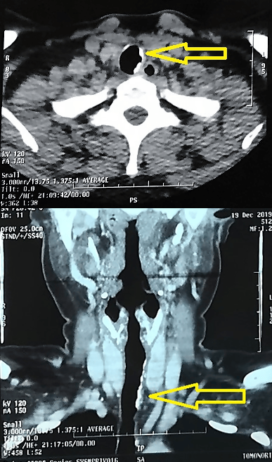
Source: Provided by the authors from imaging examinations of the case studied.