Reporte de caso
← vista completaPublicado el 24 de febrero de 2025 | http://doi.org/10.5867/medwave.2025.01.3012
Comunicación oronasal de la línea media del paladar inducida por cocaína: reporte de caso
Cocaine-induced oronasal communication of the midline of the palate: A case report
Abstract
Cocaine abuse poses a significant public health challenge, leading to severe systemic and localized complications. Intranasal cocaine use can result in chronic rhinitis, septal perforation, and palatal perforation due to its vasoconstrictive effects, which cause ischemia and tissue necrosis. We present the case of a 44- year-old woman with a 10-month history of palatal perforation, attributed to 12 years of chronic cocaine use, presented with nasal regurgitation, feeding difficulties, and cachexia. Examination revealed a 3 x 2 cm palatal perforation, nasal asymmetry, and a saddle nose deformity. Computerized tomography scans showed extensive nasal septum perforation and sinus mucosal thickening. Initial treatment involved antibiotics for sinusitis, followed by the fabrication of an obturator prosthesis to improve speech and feeding. The chronicity and extent of the palatal and nasal damage illustrates the severe consequences that can arise from sustained abuse. This case highlights not only the physical manifestations but also the challenges in managing such cases, emphasizing the necessity of a multidisciplinary approach. The integration of dental, otolaryngological, and psychological care is crucial for both immediate and long-term management. The main lesson from this case is the importance of comprehensive, patient-centered care that prioritizes stabilization and quality of life while supporting the patient’s path to rehabilitation. Provisional treatment with an obturator prosthesis can provide significant improvement in speech and feeding, providing a viable solution until the patient can maintain abstinence. Conservative management and prosthetic rehabilitation remain effective options, reinforcing the need for individualized, multidisciplinary care strategies.
Main messages
- Chronic cocaine abuse leads to nasal and palatine perforation, which affects vital functions.
- Cocaine-induced midline destructive lesions resemble systemic diseases. Therefore, it is essential to consider differential diagnoses.
- It is crucial to avoid consumption for six months before reconstructive surgery, and comprehensive treatment with prostheses and rehabilitation support is necessary.
Introduction
Cocaine abuse poses a significant public health challenge, leading to severe systemic and localized complications [1]. This drug inhibits neurotransmitter reuptake,producing euphoria and heightened alertness [2]. The main forms of cocaine consumption are cocaine hydrochloride and crack cocaine, with inhalation being the preferred method due to its faster and more potent effects [3].
Intranasal cocaine use can severely damage nasal and oral structures due to its vasoconstrictive and anesthetic effects, leading to ischemia and tissue necrosis. This can result in chronic rhinitis, septal perforation, and, in severe cases, palatal perforation [4,5]. Cocaine-induced midline destructive lesions are classified as a distinct clinical entity with symptoms mimicking systemic diseases, including necrotizing ulcers, septal perforation, and destruction that may extend to the palate [6].
Such lesions can extend beyond the mucosal layer to affect bone and cartilage, including the nasal turbinates and ethmoid cells. This can lead to significant complications like dysphagia and nasal regurgitation, seriously impacting quality of life [6]. Additionally, patients may experience bruxism and temporomandibular joint pain due to heightened neuromuscular activity from increased dopamine neurotransmission [7]. Gingival lesions at the application site are also common, presenting with pain, erythema, and gingival recession [7].
Diagnosing cocaine-induced midline destructive lesionsrequires a thorough clinical examination, as it resembles conditions such as granulomatosis with polyangiitis, tuberculosis, tertiary syphilis, leishmaniasis, and fungal infections, all of which can cause similar nasal and osteocartilaginous destruction [6]. Imaging studies, like contrast-enhanced computarized tomography and magnetic resonance images, are valuable for assessing mucosal lesions and bone resorption in the paranasal structures. Histopathological analysis through biopsy and cultures is critical to rule out other pathologies [6]. A diagnosis of cocaine-induced midline destructive lesions requires at least two of the following findings: septal perforation, lateral nasal wall destruction, or hard palate involvement [5].
Women may be more susceptible to complete palatal perforation and exhibit a more pronounced inflammatory response in connective, osseous, and cartilaginous tissues than men [7]. Treatment often includes obturator prostheses, which help restore speech and swallowing function [8]. However, these devices may not seal completely, leading to persistent fluid leakage from the oral to the nasal cavity. Definitive surgical solutions for reconstructing soft and hard palate defects include local flaps (e.g., lingual or pharyngeal), regional flaps (e.g., galeal or pericranial), and free revascularized flaps like the forearm flap [5].
The following case report presents a patient with oronasal communication caused by midline palatal perforation induced by chronic cocaine use, highlighting the clinical manifestations, diagnostic approach, and challenges in treatment. This case report was prepared following CARE guidelines.
Case presentation
A 44-year-old woman presented at the maxillofacial surgery department with a 10-month history of palatal perforation. She reported nasal regurgitation, orofacial pain, rhinorrhea, and significant weight loss due to feeding difficulties. Her history included uncontrolled hypertension and chronic cocaine abuse for 12 years, intensified to daily use over the past five years, and a stroke in 2019. She briefly ceased cocaine but relapsed in 2021 until quitting in early 2023. She continued using marijuana daily, and the palatal lesion worsened.
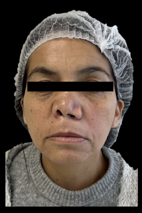
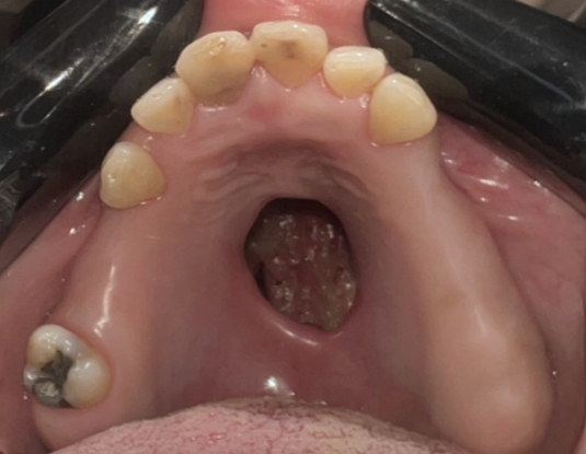
On examination, the patient appeared cachectic (body-mass index of 18.3). Facial asymmetry was noted, with nasal wing support loss leading to a saddle nose deformity marked by columellar retraction and nasal collapse (Figure 1). Intraoral findings included partial edentulism and a 3 x 2 cm irregular, deep perforation in the hard palate (Figure 2). The gums were pale pink with a scalloping loss, and the remaining teeth showed attrition and restorations without active caries.
Extraoral image of the patient.
Palatal perforation.
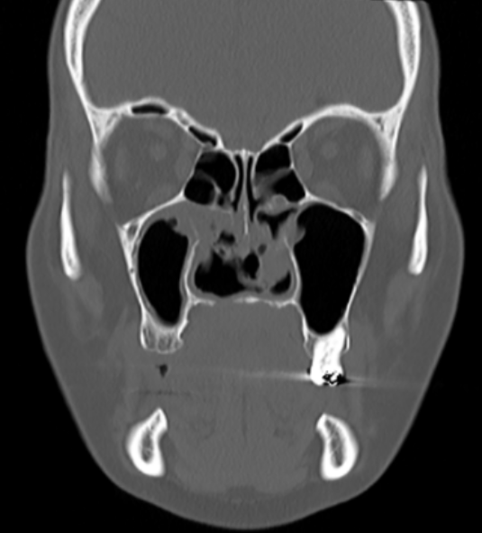
Computerized tomography scans indicated extensive nasal septum perforation involving bony and cartilaginous components, mucosal thickening in the right maxillary sinus, nasal cavity, and turbinates, with a mucous retention cyst in the left middle meatus (Figure 3). Blood analysis revealed elevated C-reactive protein (11.8 mg/L), leukocytes (10.72 x 10^3/µL), and neutrophils at 8.03%.
Computed tomography in coronal view January 2023.
The patient referred sinusitis symptoms, including fever and nasal discharge, and was initially treated with amoxicillin-clavulanic acid (500/250 mg) every 12 hours for 21 days. Due to persistent symptoms, moxifloxacin 400 mg daily was prescribed for an additional 21 days, leading to clinical improvement.
A provisional treatment plan involved a removable acrylic partial denture with a palatal obturator to improve speech and feeding. Initial treatment included supragingival scaling and oral hygiene instructions. Impressions of the upper arch were taken with the additional silicone (Elite HD Silicone + Putty Soft Normal Set), and sterile gauze was used to protect the exposed nasal mucosa. The palatal obturator was then fitted, adjusted, and polished, ensuring a proper seal and no discrepancies with the defect.
Future surgical options for definitive repair are planned once sustained cocaine abstinence is confirmed.
Discussion
This case underscores the severe complications associated with chronic intranasal cocaine use, particularly the damage to oral and nasal structures, as illustrated by this patient´s oronasal fistula. The 10-month history of palatal perforation in this case highlights the extensive harm that prolonged cocaine abuse can cause, including tissue necrosis and structural defects.
Cocaine´s primary vasoconstrictive mechanism, mediated by the inhibition of norepinephrine reuptake, results in chronic ischemia and leads to chronic ischemia, which promotes mucosal necrosis and progressive tissue damage [9]. In this case, these processes likely contributed to the extensive osseocartilaginous damage, including the hard palate and nasal septum perforation. Imaging findings of mucosal thickening and sinus cysts further supported the presence of chronic inflammation, exacerbated by ongoing cocaine use.
Palmero-Sanchez [10] emphasized the importance of thorough medical histories for diagnosing such cases, underscoring the need to document cocaine use and rule out other pathologies, such as vascular anomalies, tumors, and infections.
Oral conditions associated with long-term substance use include high caries incidence, xerostomia, salivary flow changes, enamel erosion, and gingival damage [11]. Altered olfactory function and chronic sinusitis may also indicate substance use, as observed in this patient.
Females seem to be at higher risk for cocaine-induced hard palate perforation, as reported by Cintra [12], who found findings that lower blood cocaine levels in women may lead to more intense consumption, predisposing them to significant tissue damage [9]. Women also exhibit heightened inflammatory responses, contributing to bone and cartilage damage [10]. While the typical age for such cases is around 30 years [9], this patient, at 44, demonstrates that complications can occur outside typical age ranges.
Cintra´s previous case descriptions [12] involved women with cocaine-induced palatal defects developing over approximately six years of use, with a gradual progression of the defect. By contrast, these lesions appeared after 12 years of use but exhibited rapid onset and growth. Silvestre [9] highlights the role of use frequency in tissue destruction and notes that combined cocaine and tobacco use accelerates necrosis.
The most frequently reported symptoms include rhinolalia, mucopurulent rhinorrhea, nasal ala collapse, and the loss of maxillary structures, including palatal processes and nasal turbinates [13]. This case aligns with these manifestations, with significant hard palate damage evident.
Cocaine users often suffer from head and neck complications, including paranasal sinus destruction, which predisposes them to sinusitis due to an impaired immune response [14]. This patient’s sinusitis, diagnosed after sinus wall damage, required a multidisciplinary approach with otolaryngology support. Moxifloxacin (400 mg for 21 days) effectively managed the condition.
Trimachi [15] reports that up to 88% of patients with cocaine-induced midline destructive lesions show Staphylococcus aureus colonization due to reduced oxygenation in the nasal mucosa, which promotes anaerobic pathogen growth. Such colonization complicates healing and may require targeted antimicrobial therapy.
Patient management is complicated by low adherence to treatment, often seen in those with substance abuse histories [6]. This patient's intermittent compliance posed challenges, as lesion size changes necessitated adjustments to the prosthetic obturator. Optimal treatment ideally involves surgical reconstruction after at least six months of verified cocaine abstinence, confirmed via urine metabolite testing [9].
Reconstruction considerations depend on factors such as lesion location and size, addiction duration, and infection presence. Local and regional tissue transfers in cocaine users often fail due to insufficient tissue volume and poor outcomes in central palatal defects requiring extensive access through maxillary structures [8]. A high failure rate in surgical reconstructions is associated with relapse. In this case, uncertainty regarding abstinence led to deferring surgical intervention, opting instead for an acrylic prosthesis with a palatal obturator to improve feeding and phonation while abstinence was confirmed.
Cannabis use, documented in this patient, may worsen oral health deterioration due to its association with xerostomia and increased caries risk [16]. Combined with cocaine´s effects, cannabis use may further damage mucosal and periodontal health. Additionally, the patient’s history of stroke adds complexity, suggesting multifactorial contributions to her condition.
Literature supports using obturator prostheses to rehabilitate palatal defects and improve speech and swallowing. However, persistent cocaine use can lead to necrotic margin changes, altering prosthesis fit and leading to continued rhinolalia and nasal regurgitation [5]. Thus, a multidisciplinary approach involving addiction support and social services is essential to enhance treatment adherence and outcomes. Empathetic communication and strong therapeutic alliances can foster compliance, improving prognosis and quality of life.
Silvestre´s systematic review [9], which encompasses 27 cases, and Araujo´s overview [11], which includes 22 cases, endorse conservative management with antibiotics, saline rinses, and obturators during rehabilitation, with surgery considered only after abstinence is confirmed. This approach aligns with ours, using an obturator and antibiotic therapy to manage the defect and ensure abstinence before considering reconstruction.
Conclusions
Cocaine abuse has been associated with various destructive lesions in the maxillofacial region, from nasal septum to lateral wall destruction of the nasal cavities, and, in severe cases hard palate perforation and necrosis. Accurate diagnosis requires a comprehensive clinical examination and appropriate diagnostic tests. Management options for such lesions include an acrylic partial prosthesis with an obturator or reconstructive surgery. It is essential to ensure that specific criteria are met before undertaking definitive surgical interventions for these patients.
