Reporte de caso
← vista completaPublicado el 3 de noviembre de 2025 | http://doi.org/10.5867/medwave.2025.10.3087
Displasia tanatofórica: reporte de caso
Thanatophoric dysplasia: A case report
Main messages
- Thanatophoric dysplasia is a rare and lethal genetic disease, diagnosed mainly by prenatal ultrasound and clinical findings.
- This is the first documented case in the province of Loja, highlighting the lack of official records in Ecuador.
- The report highlights limitations in access to genetic testing and comprehensive care during pregnancy.
- A possible link with pesticide exposure is suggested, an issue that requires further research in agricultural contexts.
Introduction
Thanatophoric dysplasia, or thanatophoric dwarfism, belongs to the group of fetal skeletal dysplasias. With an incidence of 1 in 10 000 to 35 000 live births, it predominates in males at a ratio of 2 to 1. It is caused by a mutation in the fibroblast growth factor receptor 3 gene, located on chromosome 4p16.3. It manifests in newborns with macrocephaly, a wide anterior fontanelle, a prominent forehead, severe hypoplasia of the midface region, and proptosis. It is also expressed with a narrow, bell-shaped chest, micromelic limbs, and redundant skin folds. The hands often present brachydactyly and a trident configuration [1,2].
There are two clinical forms of thanatophoric dysplasia: type I and type II, classified based on typical bone lesions. Type I presents with bowed femurs shaped like telephone receivers. Type II presents with a cloverleaf skull, short and straight bones, along with the findings of type I. During pregnancy, both forms may be associated with severe polyhydramnios in the third trimester [1,2].
Central nervous system abnormalities may include temporal lobe dysplasia, hydrocephalus, and lesions due to critical stenosis of the occipital foramen. Cardiac and renal abnormalities, as well as seizures, have been reported in rare cases.
Its autosomal dominant etiology involves disorganization of the growth cartilage and persistence of parenchymal tissue.
The diagnosis can be made by prenatal ultrasound from the 15th week of gestation. From the 24th week, it is possible to identify curvatures in the limbs, allowing a detection rate of 90 to 95% [3].
Two-dimensional ultrasound reveals bone lesions, while three-dimensional ultrasound allows for a more detailed visualization of the fetal face and thoracic hypoplasia [3].
The prognosis is poor, with death usually occurring in utero or shortly after birth. This outcome is generally due to respiratory failure, pulmonary hypoplasia, or compression of the spinal cord or brainstem [3].
In Ecuador, there are no official statistics reflecting the situation of thanatophoric dysplasia, making it necessary to establish national registries.
Case report
Full-term newborn, born to a teenage first-time mother, with a family history of bone dysplasia due to positional deformity and cerebral palsy due to asphyxia.
One year before pregnancy, the parents were exposed to glyphosate pesticides during their agricultural activities, without protective measures. During the first trimester of pregnancy, the mother was exposed to glyphosate herbicides without protection. She also had genitourinary infections in the last trimester, which were treated with antibiotics.
The mother attended seven prenatal checkups, undergoing five ultrasounds, the first at 6 weeks of gestation and the second at 10 weeks (Figure 1), both of which were normal. The following ultrasounds reported difficulty in observing fetal limbs.
Ultrasound at 6 weeks of gestation, with no apparent pathologies.
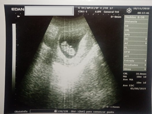
At 32 weeks, the ultrasound showed signs of bone hypoplasia, macrocephaly, facial asymmetry, with increased soft tissue in the cheeks, and short limbs. The diagnosis is consistent with thanatophoric dysplasia.
At 38.1 weeks, according to the date of her last menstrual period, the mother was admitted to the hospital in the city of Loja, Ecuador, for induction of labor due to high risk of morbidity and mortality. At that time, the ultrasound reported cranial deformation with increased biparietal diameter (10.9 centimeters), non-visible facial bones, femoral length of 3.5 centimeters, abdominal circumference of 31.4 centimeters, hypoplastic femurs, grade III fundal placenta with fetal heart rate of 145 beats per minute (Table 1).
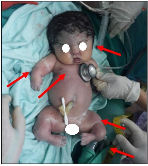
A segmental cesarean section was performed due to cephalopelvic disproportion. Meconium-stained amniotic fluid was identified. A live female newborn was delivered with an Apgar score of 3-1. Her gestational age was 38 weeks (Capurro), with a weight of 2370 grams (10th to 50th percentile), head circumference of 34 centimeters (3rd to 10th percentile), height of 36 centimeters (10th to 50th percentile), chest circumference of 27.5 centimeters, heart rate of 45 beats per minute, apnea, and temperature of 36 degrees Celsius. The newborn was hypoactive, cyanotic, not crying, and had a capillary refill time greater than 3 seconds. In addition, she presented with craniotabes, macrocephaly, with wide and tense fontanelles, and separation of the cranial sutures. She also had exophthalmos, bilateral corneal opacity, absence of pupillary reflex, and a wide nasal bridge with greenish-yellow discharge.
The auricles were typically implanted, the oral cavity was small with two incisors, and the soft palate was intact. In addition, the neck was short, the thorax was short and symmetrical with no respiratory movements, no pulmonary murmur, and bradycardia.
The abdomen was soft with visceromegaly, hydroaerial noises were present, and rectus diastasis was noted. The umbilical cord was thick, with two arteries and one vein. The female external genitalia were normal, and the anal region was permeable. The limbs showed micromelia, absent tone and strength, and no primary reflexes (Figure 2).
Phenotypic features of thanatophoric dysplasia in the newborn.
Resuscitation maneuvers (stimulation, suction, oxygen via mask) were performed without response, and palliative care was then initiated. The girl died 15 minutes after birth.
The diagnosis was a full-term newborn, low birth weight for gestational age, severe neonatal asphyxia, and congenital malformation of the thanatophoric dysplasia type. The family did not accept genetic counseling or an autopsy.
Discussion
Thanatophoric dysplasia is diagnosed based on clinical, morphological, radiological, and genetic criteria. Molecular confirmation requires identification of a mutation in the FGFR3 gene. This procedure is limited in Ecuador and neighboring countries, forcing healthcare professionals to rely mainly on ultrasound and phenotypic findings [4].
This case is the first documented report of thanatophoric dysplasia in the province of Loja in Ecuador. There was no evidence of specialized follow-up with confirmatory prenatal molecular studies (genetic analysis) or postmortem studies (autopsy). Consequently, the diagnosis was based exclusively on clinical and morphological findings.
This case reflects economic limitations, deficiencies in healthcare infrastructure, sociocultural barriers, and the absence of public policies. All of these shortcomings are factors that affect access to dignified and timely health care.
Other similar situations have been reported in the country. For example, at the Isidro Ayora Gynecology and Obstetrics Hospital in Quito, there was a case of thanatophoric dysplasia in a 23-year-old pregnant woman with no relevant obstetric history. At 29 weeks of gestation, ultrasound revealed short limbs and kyphoscoliosis. Therefore, fetal karyotyping was performed without molecular genetic studies. The newborn died one minute after birth, and the autopsy reported classic signs of thanatophoric dysplasia, including a narrow chest, severe micromelia, pterygium colli, and hypertelorism [5].
In Ecuador, the lack of official records of bone dysplasias by the National Institute of Statistics and Census makes it difficult to determine the incidence of these diseases. This limits the development of public policies aimed at their detection and management.
Experiences in countries such as Colombia, Mexico, and Spain report cases of micromelia, funnel chest, macrocephaly, and pulmonary hypoplasia, with fatal perinatal outcomes. However, in these contexts, genetic studies, 3D ultrasounds, and autopsies are more frequently available. This allows for confirmation of the diagnosis and enrichment of epidemiological records [6,7,8].
A study conducted in Ecuador with 26 pregnant women identified high concentrations of pesticide metabolites (dialkyl phosphates and ethylenethiourea, derivatives of dithiocarbamates), even in women with no connection to farming. No cases of bone dysplasia were reported in this study [9]. However, a possible association between exposure to pesticides (glyphosate) and the development of congenital malformations is suggested.
A multicenter study based on data from the National Birth Defects Prevention Study (1997 to 2002) analyzed the association between maternal occupational exposure to herbicides during the periconceptional period and congenital malformations. Although this study did not find a significant relationship with specific skeletal dysplasias, it did report an association with gastroschisis in women over 20 years of age with combined exposure to multiple pesticides [10].
Conclusions
The lessons learned in this case provide valuable guidance for addressing future cases in our setting. Thanatophoric dysplasia is a lethal skeletal condition. Therefore, it is essential to make an early prenatal diagnosis, integrating personal, family, and occupational history with ultrasound studies starting from the 15th week of gestation. All of these are key elements in identifying the structural alterations characteristic of this pathology.
Ultrasound monitoring during pregnancy allows for an initial diagnosis, as well as appropriate therapeutic counseling with a clear explanation of the etiology, prognosis, and management options, including the possibility of termination of pregnancy. Genetic testing should be promoted for timely diagnosis. In the case studied, the refusal to perform genetic studies or an autopsy due to stigma and religious beliefs limited the diagnostic confirmation.
Finally, although the studies reviewed do not establish a conclusive association between glyphosate and thanatophoric dysplasia, there is a need for more specific research on the effects of pesticides used in Ecuador. This will provide a better understanding of their possible implications for maternal-fetal health and thus contribute to the effective prevention of congenital malformations.
