Published on 16 de enero de 2026 | http://doi.org/10.5867/medwave.2026.01.3153
Frequency of mesenteric panniculitis in an oncologic population: A multicenter comparative study with a control group
42-year-old man with grade I-II follicular NHL with Ki67: 20%. On the left, axial CT slices and PET/CT fusion show signs of mesenteric panniculitis with hyperactive lymph nodes (SUVmax 7.3), which are more active than the liver (SUVmax 2.7). The central image shows CT demonstrating increased mesenteric fat density and volume. Right image: MIP of PET showing only mesenteric involvement.
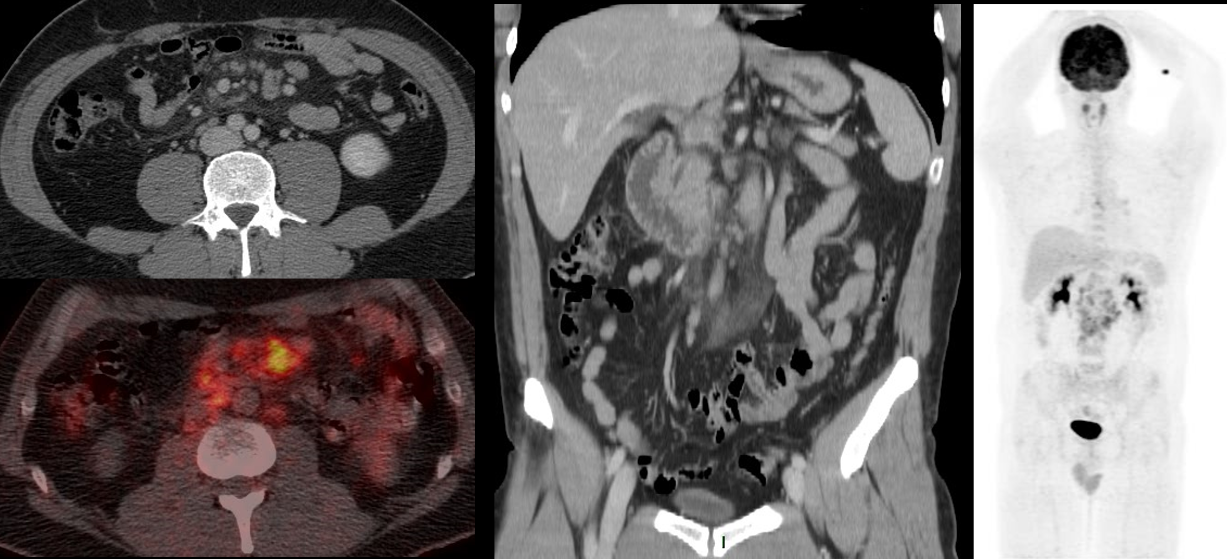
NHL: non-Hodgkin lymphoma, CT: computed tomography, PET/CT: positron emission tomography/computed tomography; SUVmax: maximum standardized uptake value, MIP: maximum intensity projection.
Source: Prepared by the authors based on a case included in the case series.
Source: Prepared by the authors based on a case included in the case series.